The pelvic diaphragm is a muscular partition formed by the Levatores Ani and Coccygei muscles and associated connective tissue, including the parietal pelvic fascia on their upper and lower aspects. In order to accommodate the birth canal, a female’s pelvic cavity is larger than a male’s. This is the reason that the pelvic floor tends to be considered a part of female anatomy, even though males have an equivalent pelvic floor.
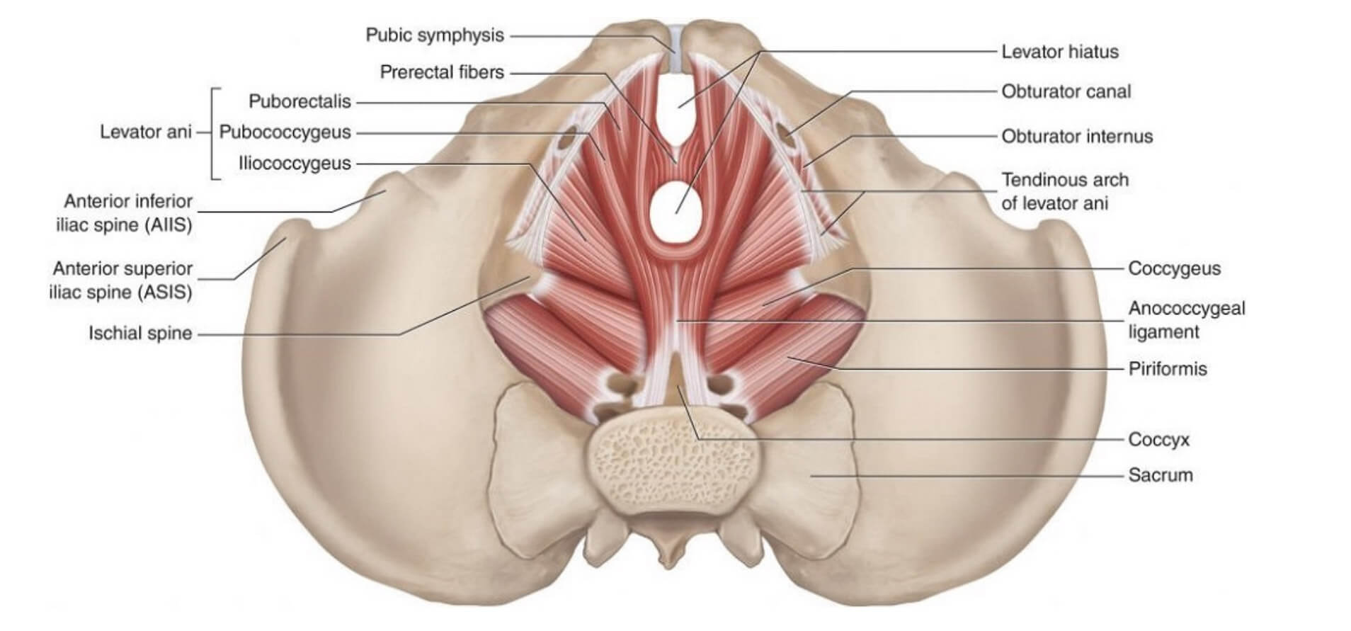
The levator ani is a broad, thin muscle group, situated on either side of the pelvis. It is formed from three muscle components:
It is anchored to the inner surface of both sides of the lesser pelvis, joining to form the majority of the pelvic floor. It supports the viscera in the pelvic cavity and surrounds the structures that pass through it. Contraction of the levator ani muscles also aid in maintaining urinary and fecal continence until voluntary voiding occurs.
ORIGIN: Posterior surface of bodies of pubic bones
INSERTION: Anococcygeal ligament, coccyx, perineal body and musculature of prostate/vagina
ACTION: Draws the distal rectum upward/forwards. Aids in contraction of the anal sphincter.
ORIGIN: Fascia of the internal obturator muscle and ischial spine
INSERTION: Anococcygeal ligament and coccyx
ACTION: Support the pelvic viscera, elevates the pelvic floor and comprises part of the pelvic diaphragm.
Puborectalis Muscle
ORIGIN: Posterior surface of bodies of pubic bones
INSERTION: None
Coccygeus is a sheet of muscle and fibrous tissue of the pelvis. Together with the levator ani, it comprises the pelvic diaphragm that forms the inferior wall of the true pelvis ( lower, narrower part of the pelvis).
Often the coccygeus acts in contact with the lower border of piriformis in several supporting actions.
ORIGIN: Ischial spine
INSERTION: Inferior end of sacrum, Coccyx
ACTION: Supports the pelvic viscera and elevates the pelvic floor. It also has a minor role in flexing the coccyx.
The pelvic floor muscles play a crucial role in supporting internal organs, maintaining continence, and contributing to overall stability. They are essential for various bodily functions, including childbirth, sexual health, and core strength. Strengthening these muscles through targeted exercises and more rehabilitative ones, such as Kegels, can help prevent and manage certain conditions like pelvic organ prolapse.
Support for Organs
Bladder and Bowel Control
Sexual Function
Core Stability
Assists in Childbirth (for Women)
Pressure Regulation
The pelvic floor muscles are primarily made up of thick skeletal muscles with nearby ligaments and their investing fascia. It has a basin-shaped muscular diaphragm and works with the transversus abdominis and the lumbar multifidus to stabilize the pelvis. Its main function is to stabilize the bottom of the abdominal cavity to help support the inferior organs of the pelvis, such as the bladder, prostate, uterus and rectum.
For postural support, the most important muscle of the pelvic floor is the levator ani, which is composed of three different muscle units: the pubococcygeus, iliococcygeus and puborectalis. These muscles form a cup or a diaphragm that has the capacity to contract in and up.
Histologically, the majority of the pelvic floor muscles are made up of slow-twitch or type I muscle fibers. Recall that type I fibers are ideal for long periods of contraction, while type II fibers are needed for a quick response to physiological changes. It is important to maintain a correct and synergistically working correlation between the pelvic floor and the rest of the Inner Unit of the Core.
Strength and balance in the pelvic floor are greatly influenced by pelvic orientation—whether in neutral, anterior, or posterior positions. In everyday tasks, it’s essential to evenly recruit the pelvic floor muscles for optimal function. To ensure proper recruitment and alignment, training with specific exercises, such as those provided by KinetiCode®, helps promote the correct pelvic orientation.
This approach is crucial not just for specific events like childbirth, but for maintaining pelvic floor health throughout daily activities and sports.
The pelvic floor plays a vital role in providing structural support to the pelvic organs. However, in many surgeries such as hernia repairs, hysterectomies, and Caesarean childbirth, the Inner Unit muscles are often cut, which reduces communication with the pelvic floor. As a result, any dysfunction in these muscles can lead to instability of the pelvic organs, such as prolapse, and incontinence.
Pelvic floor disorders are more commonly encountered in females than in males, primarily due to factors like obstetric causes and hormone-related ligament laxity, which occur exclusively in females. Consequently, organ prolapse remains largely a gynecological problem.
To address pelvic floor dysfunctions, specific KinetiCode® exercise programs are designed to tighten and tone the pelvic floor muscles while also re-establishing the connection between the pelvic floor and the rest of the Inner Unit muscles of the Core.
Additionally, one of the most common interventions for women during pregnancy or after childbirth is the practice of Kegel exercises (also known as pelvic floor exercises). These exercises aim to isolate and train the pelvic floor muscles, helping to prevent or stop urinary incontinence.
The Core Structure & Function
Mentor Exercise Review
A Mentor Review is a live guided session where you can present exercises, ask questions, and receive individual feedback on your execution, cueing, and reasoning.
Please prepare (at least) one exercise you would like feedback on. Active participation is encouraged to gain the full benefit of the session.
Sessions require a minimum of two participants. If fewer than two students are registered 48 hours in advance, the session will be cancelled.
Mentor Reviews are scheduled based on participant availability. You will receive a confirmation email once your session is finalized.
©KINETICODE® 2025. ALL RIGHTS RESERVED
The Spine & Neck – Back Muscles Lectures – Part 2
©KINETICODE® 2026. ALL RIGHTS RESERVED
The Spine & Neck – Back Muscles Lectures – Part 1
©KINETICODE® 2026. ALL RIGHTS RESERVED
The Pelvis & Posterior Abdominal Wall Lectures –
Part 2
©KINETICODE® 2026. ALL RIGHTS RESERVED
The Pelvis & Posterior Abdominal Wall Lectures –
Part 1
©KINETICODE® 2026. ALL RIGHTS RESERVED
The Core Structure & Function Lectures – Part 2
©KINETICODE® 2026. ALL RIGHTS RESERVED
The Core Structure & Function Lectures – Part 1
©KINETICODE® 2026. ALL RIGHTS RESERVED
©KINETICODE® 2026. ALL RIGHTS RESERVED
Price of the Course: €2.500
(excl. VAT, reclaimable)
*Prefer a payment plan? Email us at academy@kcmove.nl with a proposal.
Invest in your future as a Pilates professional with Europe-wide recognized certification. This fee gives you full access to a carefully designed 7-month learning journey that blends science, practice, and mentorship.
Your investment includes:
©KINETICODE® 2026. ALL RIGHTS RESERVED
The KinetiCode® Pilates Certification follows a 7-month study cycle, designed for maximum flexibility. You can begin at the start of any month and progress at your own pace while completing all required coursework.
In total, the course includes:
Total course time = 250 hours
©KINETICODE® 2026. ALL RIGHTS RESERVED
This website uses cookies to enhance your experience. Some are essential for site functionality, while others help us analyze and improve your usage experience. Please review your options and make your choice.
If you are under 16 years old, please ensure that you have received consent from your parent or guardian for any non-essential cookies.
Your privacy is important to us. You can adjust your cookie settings at any time. For more information about how we use data, please read our privacy policy. You may change your preferences at any time by clicking on the settings button below.
Note that if you choose to disable some types of cookies, it may impact your experience of the site and the services we are able to offer.
Some required resources have been blocked, which can affect third-party services and may cause the site to not function properly.